





Advanced Retinal Imaging: Detecting Glaucoma and Macular Degeneration Without Dilation
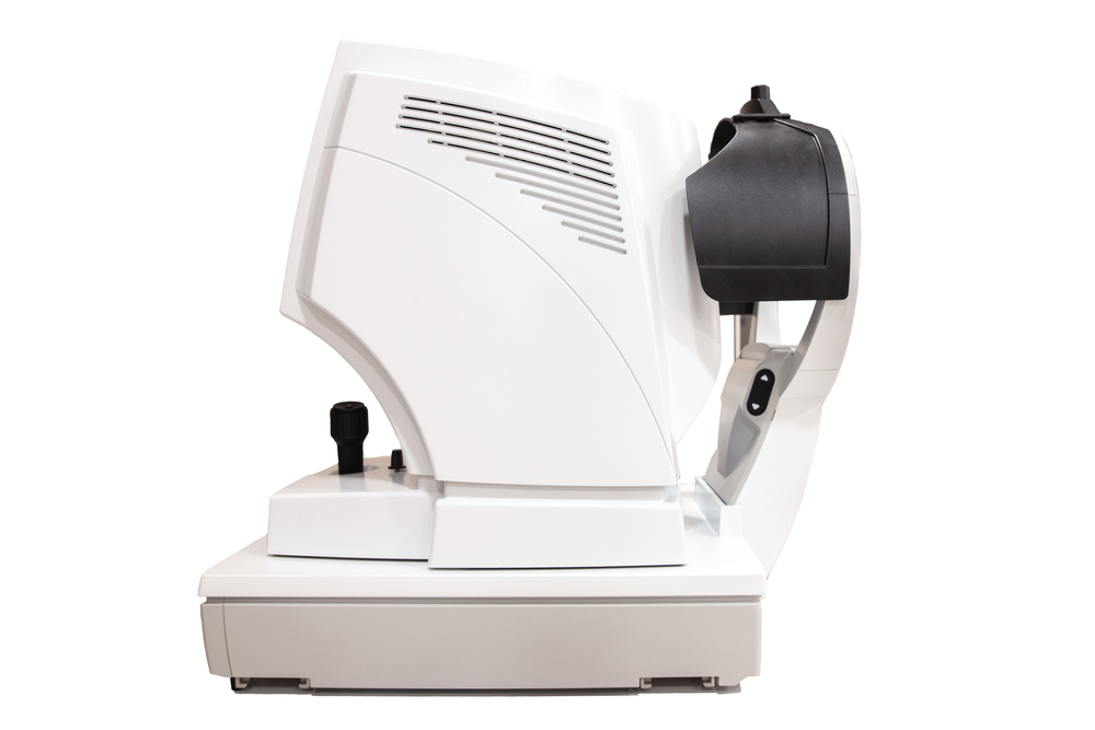
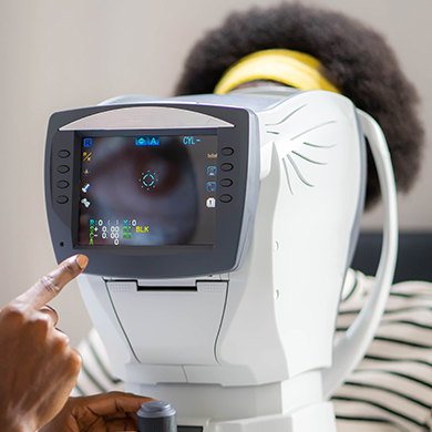
Many serious eye diseases can begin developing before you notice changes in your vision. At Visual Health Doctors of Optometry, we use advanced retinal imaging to get a detailed look at the structures inside your eyes and identify subtle changes that may require closer attention. Modern imaging technology can capture valuable information about the retina, macula, and optic nerve quickly and comfortably, often without requiring dilation for the imaging itself.
What Is Advanced Retinal Imaging?
Retinal imaging uses specialized technology to create detailed images of the back of the eye. Digital retinal photography captures high-resolution views of the retina, blood vessels, macula, and optic nerve. Optical coherence tomography, or OCT, provides detailed cross-sectional images that allow us to examine layers of retinal and optic nerve tissue.
These images also become part of your eye health record. By comparing scans from one visit to the next, we can look for subtle changes that may not yet be affecting your day-to-day vision.
Detecting Glaucoma Before Vision Loss Occurs
Glaucoma can gradually damage the optic nerve, and early stages may cause few noticeable symptoms. Advanced eye imaging helps us evaluate the optic nerve for changes that may indicate glaucoma or an increased risk for the disease.
During a comprehensive eye exam, we may use imaging alongside other glaucoma testing to evaluate factors such as:
- Changes in the appearance of the optic nerve
- Thinning of nerve fiber tissue
- Differences between the two eyes
- Progressive changes documented between examinations
- Other abnormalities that may require additional glaucoma testing
Identifying Signs of Macular Degeneration
Age-related macular degeneration affects the macula, the central portion of the retina responsible for detailed central vision. Because changes can develop before noticeable vision loss, detailed retinal imaging can be especially useful for monitoring the macula.
OCT imaging allows us to examine retinal layers closely and look for structural changes associated with macular disease. Retinal photography can also document visible changes and create a baseline that we can compare with future exams. Patients with a personal or family history of macular degeneration may particularly benefit from regular monitoring.
Does Retinal Imaging Mean You Never Need Dilation?
Not necessarily. One major advantage of modern retinal imaging is that detailed images can often be captured without dilating your pupils first. This can make the imaging process convenient and comfortable.
However, retinal imaging and dilation provide different information. Depending on your symptoms, medical history, risk factors, or what we see during your exam, we may still recommend a dilated eye examination. Advanced imaging is an important diagnostic tool, but it does not eliminate the need for dilation in every patient.
Contact Visual Health Doctors of Optometry to schedule your comprehensive eye exam and retinal imaging today. Visit any of our offices in Alexandria, Arlington, Ashburn, Burke, Fairfax, Falls Church, Herndon, or Woodbridge, Virginia. Contact a location nearest you, or book an appointment on our website.


© 2026 Visual Health Doctors of Optometry.
Eye Doctor & Optometrist in Alexandria, VA, Arlington, VA, Ashburn, VA, Burke, VA, Fairfax, VA, Falls Church VA, Herndon, VA, Mclean, VA, Reston, VA & Woodbridge, VA
Eye Doctor & Optometrist in Alexandria, VA, Arlington, VA, Ashburn, VA, Burke, VA, Fairfax, VA, Falls Church VA, Herndon, VA, Mclean, VA, Reston, VA & Woodbridge, VA
Powered by: 